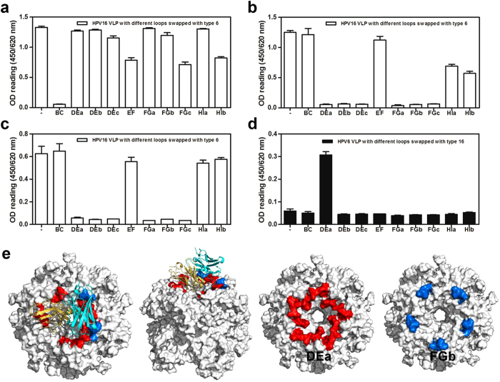
Figure 3. Identification of critical loops of the L1 protein for mAbs binding.
Binding of a murine N-mAb (1A4) with BC loop specificity (a), H16.V5 (b) and 26D1 (c) to VLPs with HPV16 wild-type and HPV16 hybrid VLP mutants. VLP 16 wild-type (“-” on X-axis), HPV16 VLP with different loops swapped with the type 6 loops displayed as open bar. (d) Antibody 26D1 was tested by ELISA for reactivity to HPV6 wild-type and HPV6 hybrid VLP mutant. VLP 6 wild-type (“-” on X-axis), HPV6 VLP with different loops swapped with the type 16 loops displayed as solid bar. (e) Binding loop prediction by homology modeling and molecular docking. A model of 26D1 Fab is displayed as a solid ribbon diagram. The yellow and cyan ribbons represent the light chain and heavy chain, respectively. Predicted binding loops marked with red and blue are DEa loops and FGb loops. Residues involved in the 26D1 binding were mostly located at the DEa loop (red) and fewer at the FGb loop (blue).

