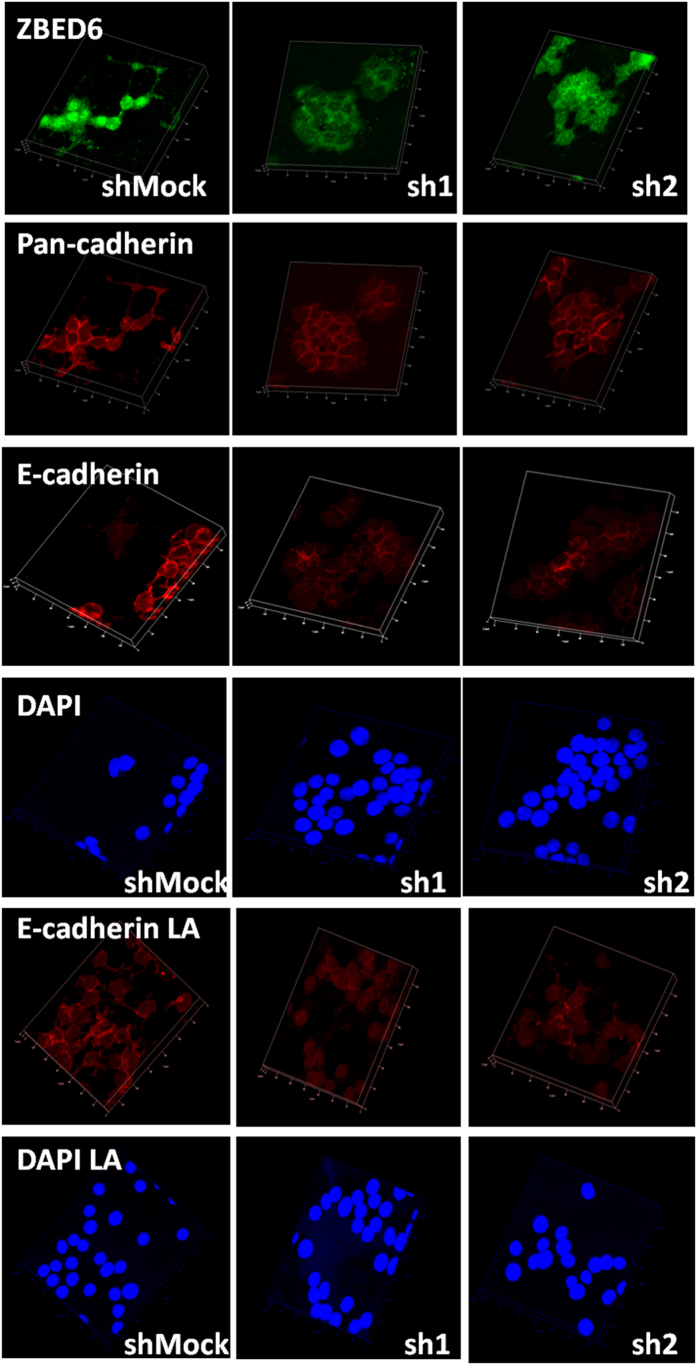
Figure 4. Staining of shMock, sh1 and sh2 βTC6 cells with ZBED6, pan-cadherin and E-cadherin antibodies.
Equal numbers of cells were seeded onto cover slips with or without 10 μg/ml mouse laminin (LA) coating. After 3 days culture, cells were fixed and stained. Images were generated from confocal Z-stack scanning using Imaris 3D model. Results are representative for 3 independent experiments. The size of one unit of the XY frame is 20 μm.

