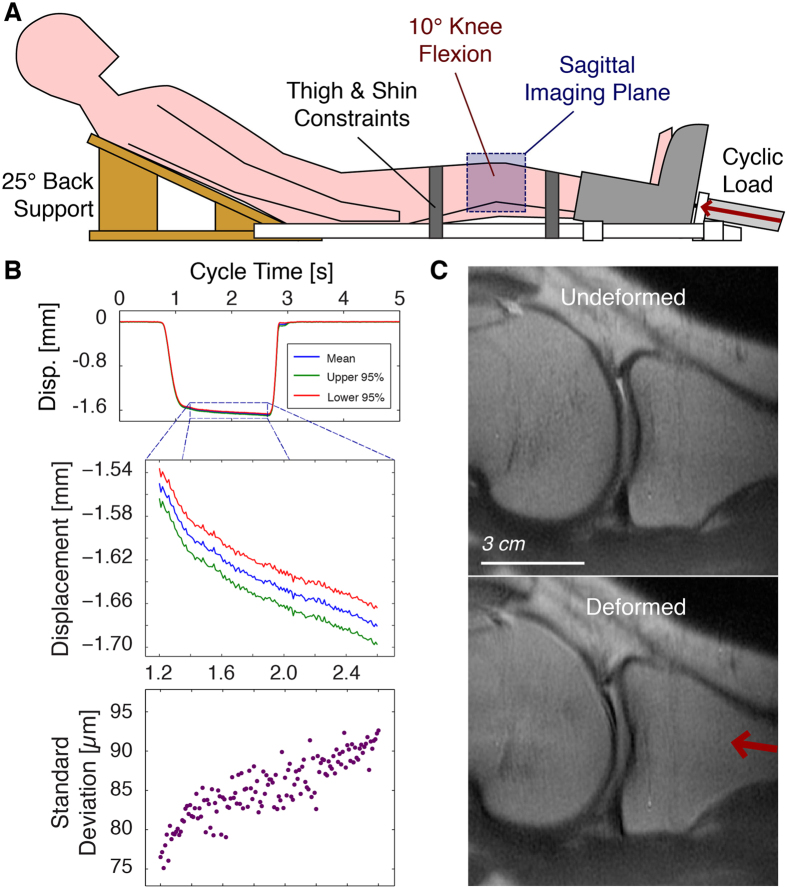
Figure 2. MRI-compatible knee loading device for in vivo measurement of tissue deformation.
A loading device was manufactured to permit the cyclic loading of the leg of a human subject by a pneumatic actuator within the confines of a clinical MRI system (A). A laser displacement system, used only outside of the MRI, allowed for the measurement of leg motion across multiple loading cycles (B). Straps across the thigh and shin were used to restrict concomitant motion of the knee (flexed at ~10°) under compression, permitting a sagittal slice through the undeformed and deformed knee tissues to be imaged during cyclic loading (C). Although the cartilage can be imaged before and during loading, the measurement of nominal measures does not provide internal information, and there is insufficient image texture for digital image correlation techniques. Because dualMRI is based on phase contrast, internal displacements can be measured at each imaged pixel, providing intratissue deformations not otherwise measurable.

