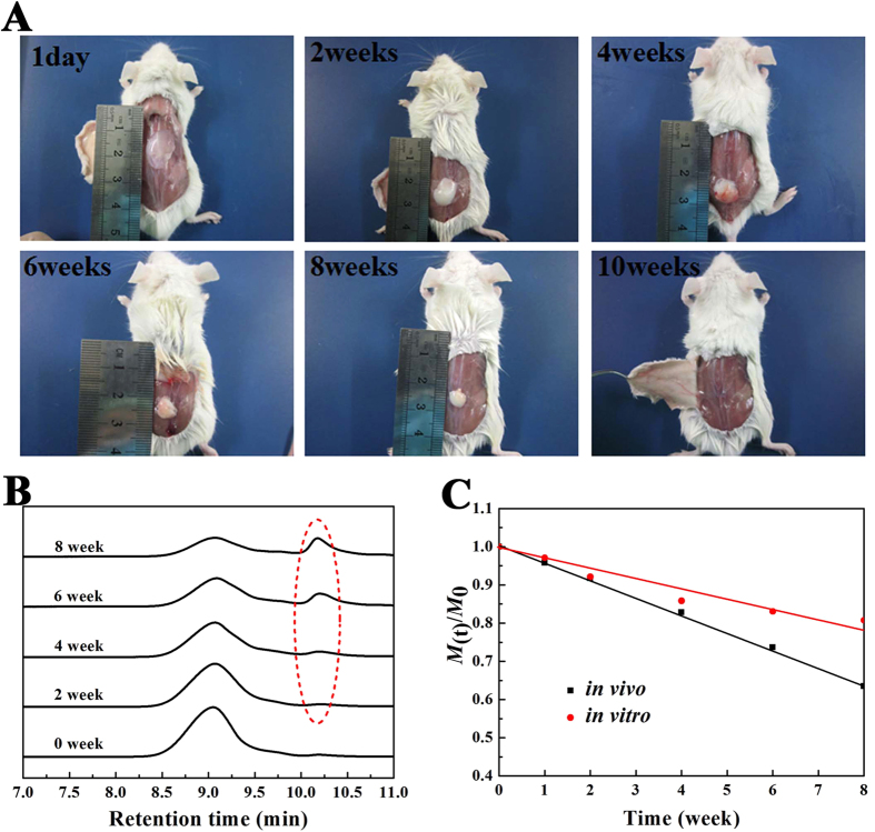
Figure 6. In vivo degradation of the hydrogels after subcutaneous injection of 25 wt% copolymer in the normal saline solution into the back of the BALB/c mice.
(A) in vivo gel maintenance. Images were taken at the pre-determined time after subcutaneous administration. The images was representative of n = 3 at each pre-determined time. The remaining hydrogel became smaller with time and completely disappeared by week 10; (B) GPC traces of PDLLA-PEG-PDLLA copolymer collected during in vivo degradation after subcutaneously injected in mice. The dashed circle emphasized the peaks of degradation products of low molecular weights; (C) Change of normalized molecular weight (M(t)/M0) of copolymers in the remaining hydrogels during in vitro and in vivo degradation. Here, M0 represented the MW of the copolymer before degradation.

