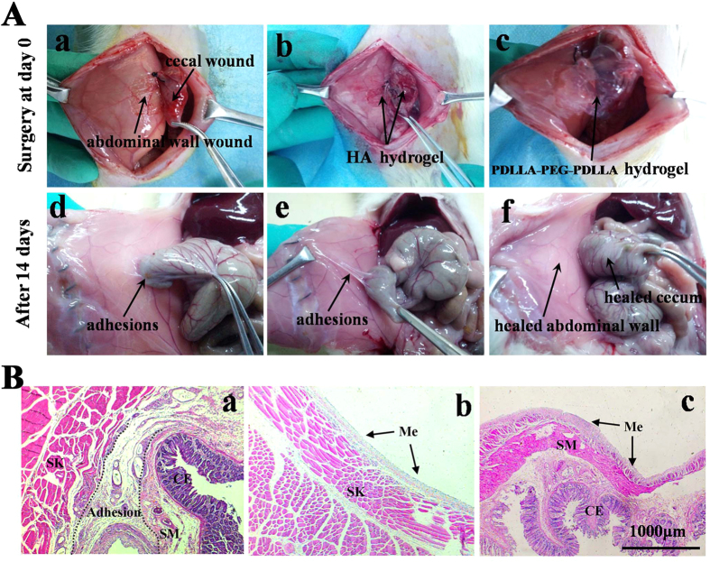
Figure 7. In vivo application and evaluation of adhesion-prevention effect in rat model.
The images was representative of n = 8. (A) Photographs of animal experiments of post-operative adhesions. (a to c) In operation; (d to f) gross observations of the efficacy of prevention adhesion after 14 days; (a and d) The untreated defect was used as the negative control group and a firm adhesion was observed 14 days after operation; (b and e) HA hydrogel applied on the injury sites and moderate adhesion was observed in (d); (c and f) The injury sites were treated by PDLLA-PEG-PDLLA hydrogel and no apparent adhesion was observed in (f), with healed abdominal wall and cecum. (B) Optical micrographs of HE-straining slices for the injured site 14 days after surgery. (a) Adhesion between the defective cecum and abdominal wall from the animal without treatment. (b and c) Healed abdominal wall (b) and cecum (c) treated with the PDLLA-PEG-PDLLA hydrogel. A thin layer of remesothelialized tissue containing a larger number of mesothelial cells formed on the defect site. ME: mesothelial cells; SK: abdominal wall skeletal muscle; SM: visceral smooth muscle; CE: cecal mucosa. Magnification: 50 ×.

