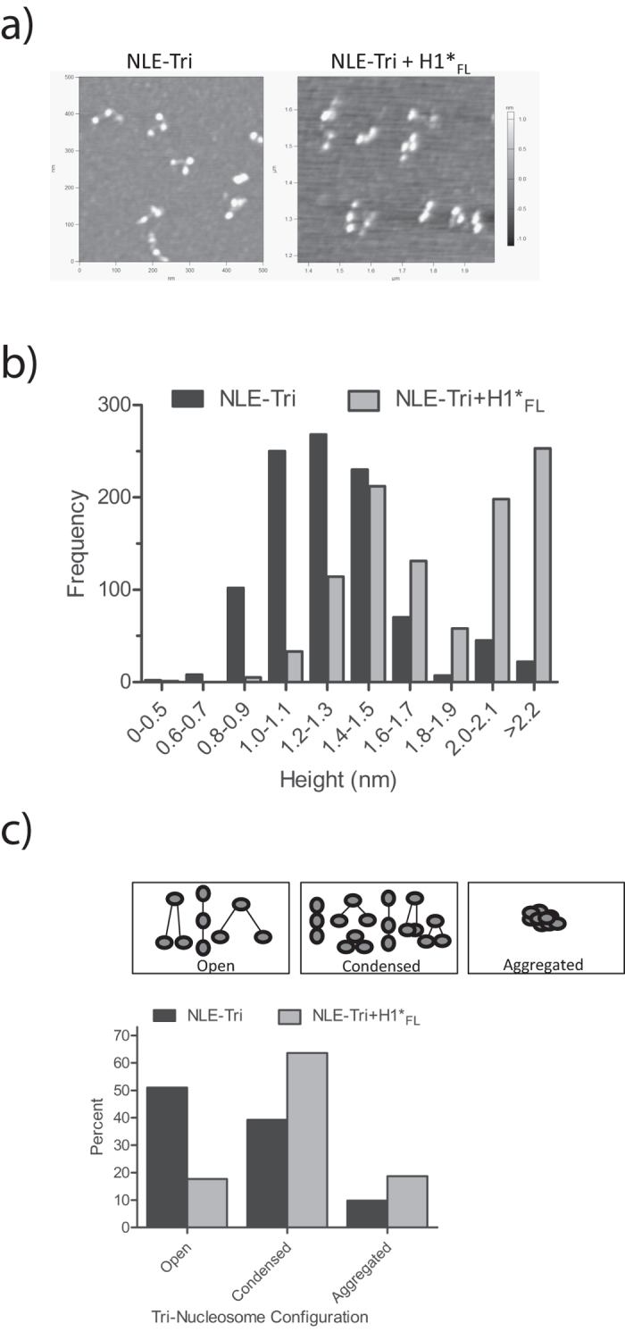
Figure 4. H1 compacts trinucleosomes.

(a) NLE-Tri was imaged by atomic force microscopy (AFM) in absence of H1FL (left), or with a 1:1 ratio of H1 per tri-nucleosome (right). (b) Binned height profiles of both NLE-Tri alone and in the presence of H1FL. A minimum of 9 separate images were used to complete height traces (Fig. S4A) on a total of 1005 nucleosomes either with or without H1FL and those heights are depicted in this graph. The mean height distribution of NLE-Tri alone is ~1.2 nm and increases to ~1.5 nm in the presence of H1FL. Importantly, the propensity for aggregates increases significantly with H1FL present. (c) Each of the observed arrangements was seen in an open conformation (longer distance between the nucleosomes - open), or in a condensed conformation with closely spaced nucleosomes. Aggregation of sample was also observed. A minimum of seven separate images for both NLE alone or with H1FL were used to count the number of trinucleosomes in each group for a total of 481 NLE-tri alone and 524 NLE-Tri with H1FL. The graph depicts the number of trinucleosomes found in each group in the absence or presence of H1FL.
