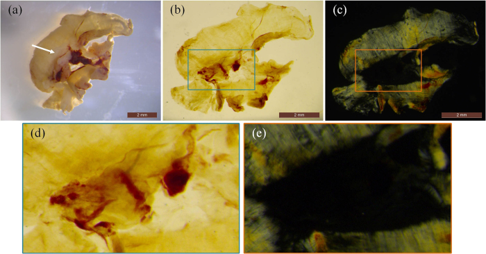
Figure 2. The muscle fibers after clearing is observed by polarized light.
(a) a photo of the diaphragm sample before the clearing process. The projection view of the cleared diaphragm captured through (b) non-polarized light and (c) polarized light. Scale bar: 2 mm. (d,e) zoomed images in the boxes of (b,c).

