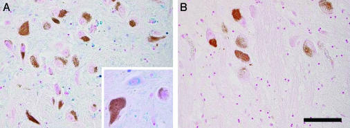
Fig. 7.
Iron histochemistry with modified Perls' staining of human SN (A) and LC (B) from a normal 88-year-old male subject. NM of dopaminergic neurons of SN and norepinephrine neurons of LC are brown granules, and iron deposits are colored in blue. (Scale bar, 100 μm.) NM-containing neurons in both SN (A) and LC (B) do not have the blue staining of iron. In SN (A), there are many iron-positive cells, which are most often oligodendrocytes with cytoplasmic iron deposits, and in LC (B) there are very few oligodendrocytes with light iron staining. Iron deposits are present also in the cytoplasm of nonpigmented neurons of SN (A) as shown at higher magnification. Iron deposits can be observed in the whole SN (A) parenchima with the exception of pigmented neurons, but they are completely absent in LC (B) parenchima.

