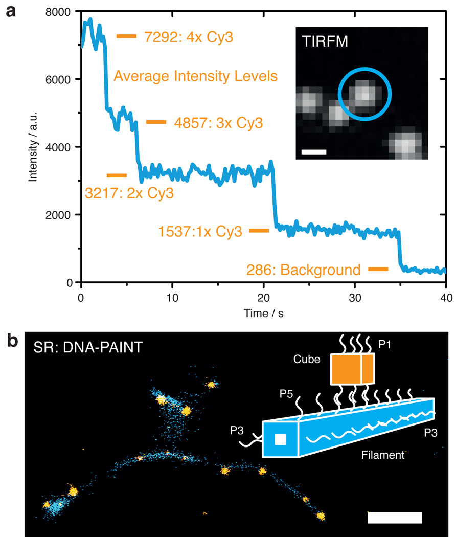
Figure 3. Fluorescence and super-resolution imaging.
a) Cubes immobilized on a surface and labeled with four Cy3 dyes show clear bleaching steps on a TIRFM setup. The inset shows a diffraction limited image of single cube structures. Length scale: 400 nm. b) Super-resolution image reconstruction of a DNA nanostructure-based complex (shown here as simplified illustration, highlighting the different binding domains P#). Cube structures (orange) hybridize to surface immobilized DNA origami filaments (blue) and are imaged using DNA-PAINT. Length scale: 350 nm.

