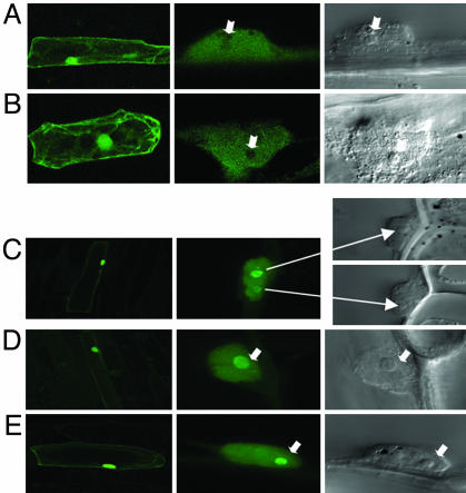
Fig. 1.
Subcellular localization of Rab17 and mRab17 in onion cells. Confocal microscopy images of onion cells transfected with Rab17-GFP (A and B) and mRab17-GFP (C-E). (Left) General views of transfected cells (×20). (Center) Detail of fluorescent cell nucleus (×60). (Right) View of the same nucleus under Nomarsky optics (arrows indicate the position of the nucleoli).

