
Figure 2. VASP ubiquitination occurs in the presence of TRIM9 and is lost following netrin treatment.
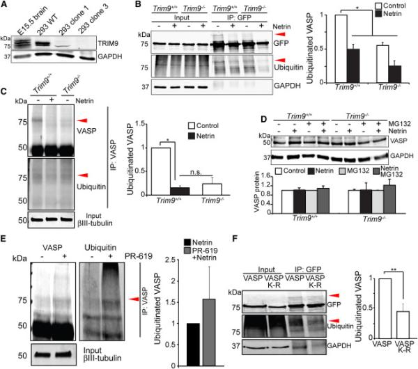
A) Immunoblot for TRIM9 and GAPDH in E15.5 brain lysates, HEK293 lysates: TRIM9+/+ (WT), TRIM9+/− (clone 1) and TRIM9−/− (clone 3). B) Input and GFP-VASP immunoprecipitation (IP) from ubiquitination assay immunoblotted (IB) for GFP, ubiquitin and GAPDH. Shown are GFP-VASP (75kDa) in TRIM9+/+ and TRIM9−/− cell lysates and ~25kDa heavier VASP band present in TRIM9+/+ lysate that co-migrates with ubiquitin (red arrowheads). Plot shows quantification of VASP ubiquitination. C) Endogenous VASP ubiquitination in TRIM9+/+ and TRIM9−/− neurons at 2DIV. A higher molecular weight VASP+ band (red arrowhead) that co-migrates with ubiquitin is seen in TRIM9+/+ cortical neurons. D) Endogenous VASP in TRIM9+/+ and TRIM9−/− control neurons or neurons treated with MG132 and/or 600 ng/ml netrin. IB for VASP (50kDa) and GAPDH. No difference in VASP protein levels detected between genotypes or treatment conditions. E) Endogenous VASP ubiquitination in TRIM9+/+ cortical neurons treated with netrin or netrin and 4 μM PR-619. Elevated ubiquitination is observed upon PR-619 treatment. F) Ubiquitination assay and quantification of wildtype VASP and VASP K-R mutant. The VASP K-R mutant exhibits a reduction in the ~25kDa heavier VASP band that co-migrates with ubiquitin (red arrowheads). All error bars represent SEM. (See also FigS2)