
Figure 5. VASP deubiquitination is required for netrin-dependent increases in filopodia density.
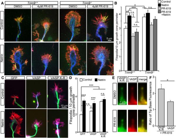
A-B) Images and quantification of filopodia +/−SEM in axonal growth cones from control, PR-619 netrin or PR-619/netrin treated TRIM9+/+ and TRIM9−/− neurons, stained for VASP (green), βIII tubulin (blue) and phalloidin (red). C-D) Images and quantification of filopodia +/−SEM in TRIM9+/+ growth cones expressing GFP, GFP-VASP or GFP-VASP K-R, stained for GFP (blue), βIII tubulin (green) and phalloidin (red). E) TRIM9+/+ filopodia containing GFP-VASP K-R (green) and mCherry-VASP (red) before and after PR-619 treatment. F) Ratio of fluorescence intensity at filopodial tip:filopodial base of GFP-VASP K-R or mCherry-VASP +/−SEM in PR-619 treated TRIM9+/+ neurons. (See also FigS5)