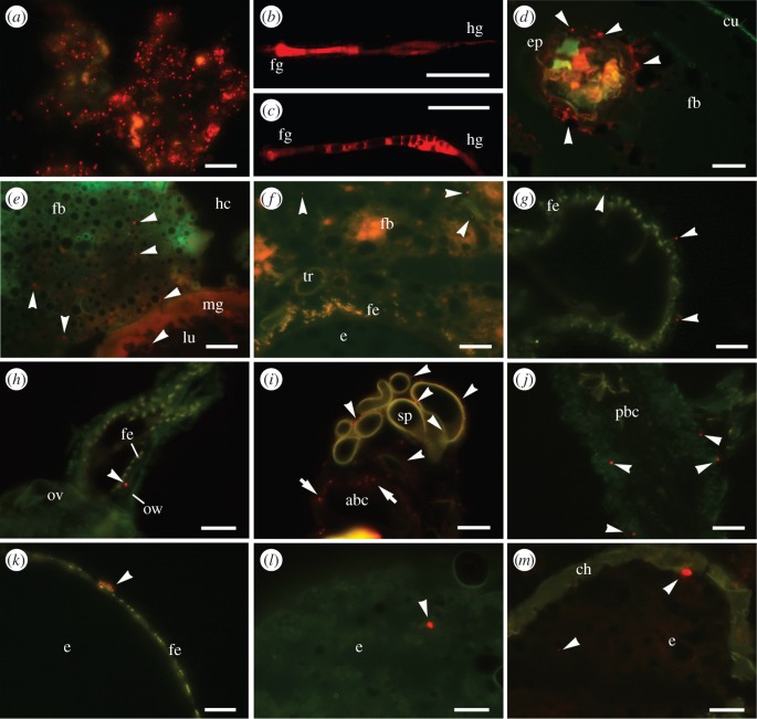
Figure 2.
Analysis of the maternal transfer of fluorescent bacteria (BioParticles®). (a) Artificial diet mixed with BioParticles® (scale bar, 50 µm). (b,c) BioParticles® in the dissected larval gut after the ingestion of the artificial diet, show (b) the foregut and midgut region and (c) the hindgut (scale bars, 1 mm). (d) BioParticles® beneath the cuticle of the larval foregut (scale bar, 100 µm). (e) Larval midgut containing translocated BioParticles® in the lumen and surrounding fat body cells (scale bar, 50 µm). (f) Female genital region with BioParticles® attached to the fat body (scale bar, 50 µm). (g,h) BioParticles® between the ovariole wall and the follicular epithelium of eggs in (g) the proximal region (scale bar, 50 µm) and (h) the distal region close to the lateral oviduct (scale bar, 150 µm). (i,j) BioParticles® associated with (i) spermatheca and the anterior bursa copulatrix, and (j) the posterior bursa copulatrix (scale bars, 50 µm). (k,l) BioParticles® attached to (k) the follicular epithelium (scale bar, 50 µm) and (l) incorporated into the yolk of dissected eggs (scale bar, 150 µm). (m) Ovipositioned egg containing BioParicles® (scale bar, 150 µm). Further details are provided in the electronic supplementary material. Arrowheads indicate fluorescent BioParticles® (red spots). abc, anterior bursa copulatrix; ch, chorion; cu, cuticle; e, egg; ep, epithelium; fb, fatbody; fe, follicular epithelium; fg, foregut; hc, haemocoel; hg, hindgut; lu midgut lumen; mg, midgut; ov, oviduct, ow ovariole wall; pbc posterior bursa copulatrix; sp, spermatheca; tr, tracheole.

