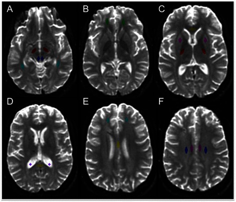
Fig. 1.

Neuroanatomic localization of regions of interest on b0 images. a Dorsolateral midbrain (blue), cerebral peduncle (maroon), and inferior longitudinal fasciculus (turquoise); b uncinate fasciculus (green); c genu of corpus callosum (yellow), posterior limb of the internal capsule (red), and putamen (pink); d splenium of the corpus callosum (yellow) and cerebrospinal fluid (purple); e anterior corona radiata (turquoise) and body of the corpus callosum (yellow); f cingulum bundle (pink) and centrum semiovale (blue)
