
Figure 1.
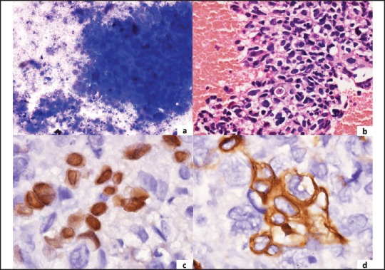
A panel of microphotographs of pulmonary squamous-cell carcinoma (a) Tumor cell cluster with necrosis in the background (MGG, ×400); (b) Cell block showing squamoid differentiation of tumor cells (H and E, ×400); (c) Nuclear positivity for p63 (IHC, ×1000) (d) Cytoplasmic positivity for CK5/6 (IHC, ×1000)