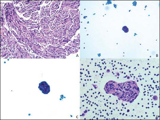
Figure 1.

(a) Spindle cells within smooth muscle fascicles. (H and E, ×200) (b) Scattered clusters of bland spindled cells in a background of histiocytes and mesothelial cells. (ThinPrep, ×200) (c) Spindle cells cluster lined by lymphatic-like endothelial cells. (ThinPrep, ×400) (d) H and E, ×400
