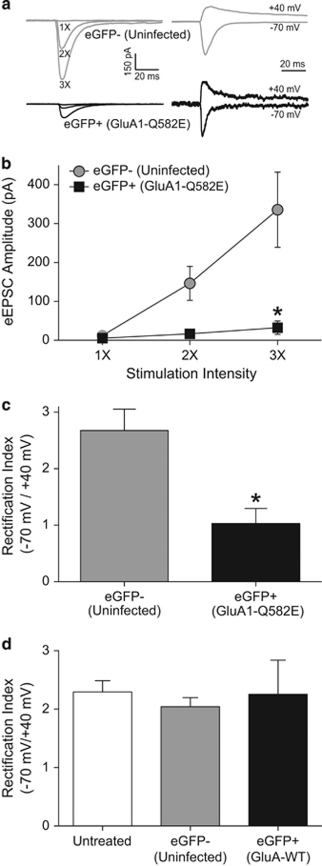
Figure 2.
The overexpression of ‘pore dead' GluA1-Q582E in accumbens shell MSNs attenuates synaptic strength and decreases inward rectification of AMPAR eEPSCs. (a) Left, representative eEPSC traces at multiples of minimal stimulation intensity in neurons expressing GluA1-Q582E (eGFP+) relative to those not expressing eGFP (eGFP−) recorded from the same slice. Right, representative eEPSC traces at +40 mV and −70 mV for eGFP+ and eGFP− neurons, used for rectification index analyses. For ease of visualization, stimulus artifacts were removed and the amplitudes were normalized to the −70 mV eEPSC in eGFP-neuron. (b) The input–output relationship of AMPAR eEPSCS illustrates decreased synaptic strength in MSNs expressing GluA1-Q582E (n=6 cells from four animals) for peak eEPSC amplitude compared with non-GFP controls (n=12 cells from six animals) at 3 × stimulation. (c) Inward rectification of AMPA eEPSCs is more prominent in eGFP− than in eGFP+ cells (ie, peak current is smaller at +40 mV relative to −70 mV). (d) Inward rectification is similar between cells from naive animals, not exposed to virus injections (untreated), cells that were not transduced by GluA1-WT injections (eGFP−), and cells overexpressing GluA1-WT (eGFP+). Untreated, nine cells from two animals; eGFP−, six cells from two animals, eGFP+, five cells from one animal.

