Figure 2.
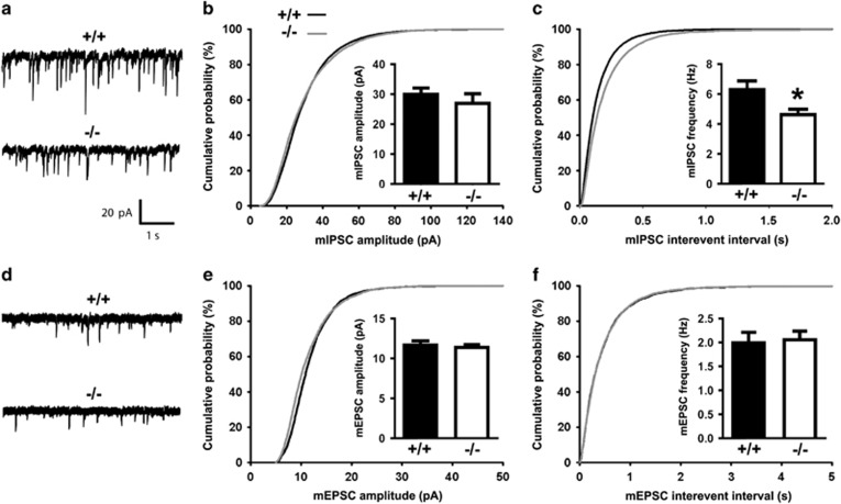
Inhibitory synaptic transmission is reduced in CA1 pyramidal neurons of Clstn2−/− mice. (a) Representative mIPSC recordings from hippocampal CA1 pyramidal neurons in acute slices from Clstn2−/− and WT adult mice. (b, c) Cumulative distributions of mIPSC amplitude (b) and inter-event intervals (c) in Clstn2−/− and WT neurons (Kolmogorov–Smirnov test, P<0.001 for inter-event intervals). Insets display mean±SEM for mIPSC amplitude (b) and frequency (c). mIPSC frequency but not amplitude was significantly reduced in Clstn2−/− neurons (Student's t-test, *P<0.05; n=10 cells for Clstn2−/− and 8 cells for WT). (d) Representative mEPSC recordings from hippocampal CA1 pyramidal neurons. (e, f) Cumulative distributions of mEPSC amplitude (e) and inter-event intervals (f) in Clstn2−/− and WT neurons. Insets display mean±SEM for mEPSC amplitude (e) and frequency (f). No significant differences were detected in mEPSCs between groups (n=8 cells for Clstn2−/− and 9 cells for WT).

