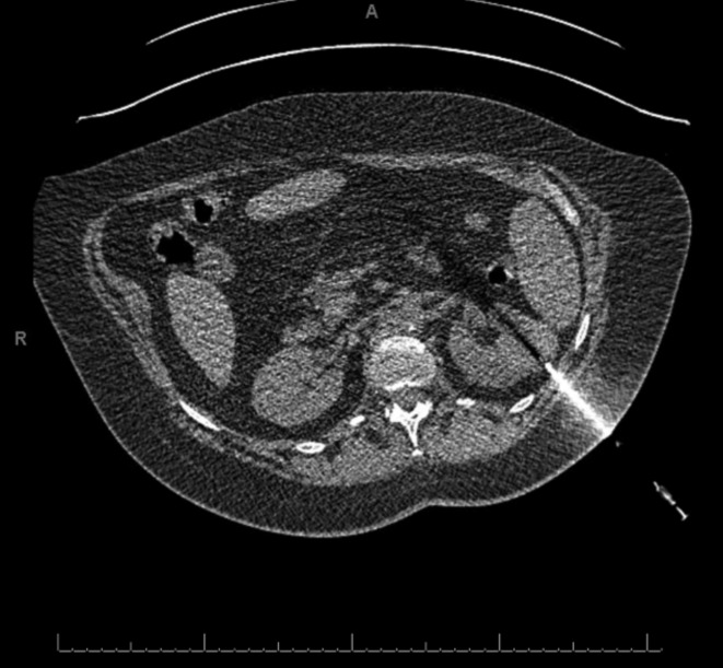
Fig. 1a.
Patient A’s CT scan during renal biopsy showing the enhancing biopsy needle being passed through the left upper pole renal mass. Patient was placed in prone position during the procedure, but the image here has been oriented for ease of comparison with Fig. 1b.

