Fig. 2b.

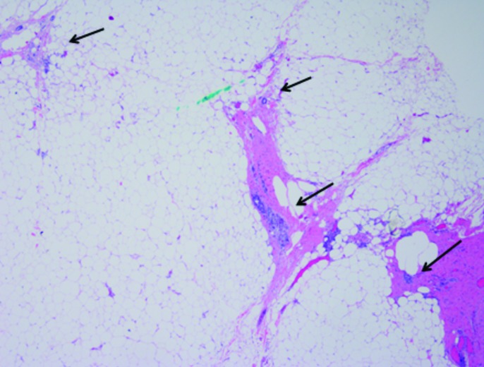
Scanning power of small, isolated, insular foci of tumour (arrows) lying in an interrupted, linear track of fibrous tissue within perinephric adipose tissue, arranged perpendicularly to the renal mass (not pictured) (20×).

Scanning power of small, isolated, insular foci of tumour (arrows) lying in an interrupted, linear track of fibrous tissue within perinephric adipose tissue, arranged perpendicularly to the renal mass (not pictured) (20×).