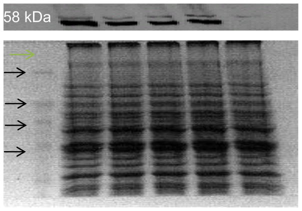
Figure 1.
Expression of human CYP1A2 in diploid yeast strains as detected by Western blots. Lanes correspond to molecular weight markers, and extracts from strains expressing CYP1A2 (YB410), CYP1A2 C406Y (YB411), CYP1A2 D348N (YB412), CYP1A2 I386F (YB413) and empty vector pRS424 (YB409), as marked. Each lane contains 10 μg of protein, as determined by Bradford assay. Equal amounts of protein were loaded as indicated by a parallel Coomassie-stained gel (bottom panel). The molecular weight markers are indicated, proceeding from 120 kDa (top), 80 kDa, 60 kDa, to 40 kDa (bottom).

