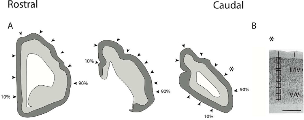
Figure 1.
(A) Illustration of a coronally-sectioned manatee brain used for neuron number estimates. In coronal planes, we systematically sampled sites (i.e., every 10th percentile) along the medial to lateral axis of the isocortex. The most medially located region is designated as the 10th percentage and the most laterally selected region is designated as the 90th percentile. Arrow-heads point to the approximate sites selected for neuron number estimates. We counted neuron numbers (layers II–VI neurons) under a unit of cortical surface area. (B) Neuron number estimates were made through the depth of the cortex. Counting frames were selected at equidistant sites through the depth of the cortex. Sites were sampled orthogonal to the axis of the cortical surface. A star shows the approximate location of (B). Scale bar is 1mm.

