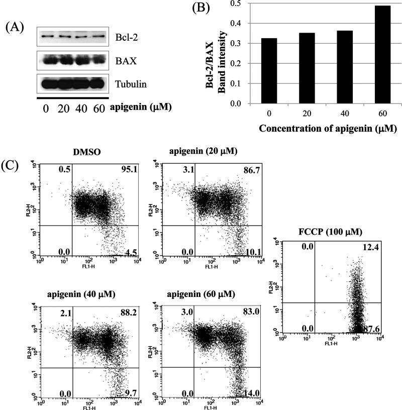
Figure 5. Apigenin induces apoptosis via extrinsic pathway in BT-474 cells.
(A) and (B) analysis of intrinsic apoptosis-related molecules. BT-474 cells were treated with apigenin (0–60 μM) for 24 h. Total proteins were analysed by western blotting with anti-Bcl-2, anti-BAX and anti-tubulin antibodies. (C) BT-474 cells were incubated with apigenin (0–60 μM) for 72 h and were dyed with JC-1 (4 μg/ml). The data were analysed by FACSCalibur flow cytometry measuring the green fluorescence and red fluorescence at 514/529 nm (FL-1) and 585/590 nm (FL-2), respectively. The data shown are representative of three independent experiments that gave similar results.

