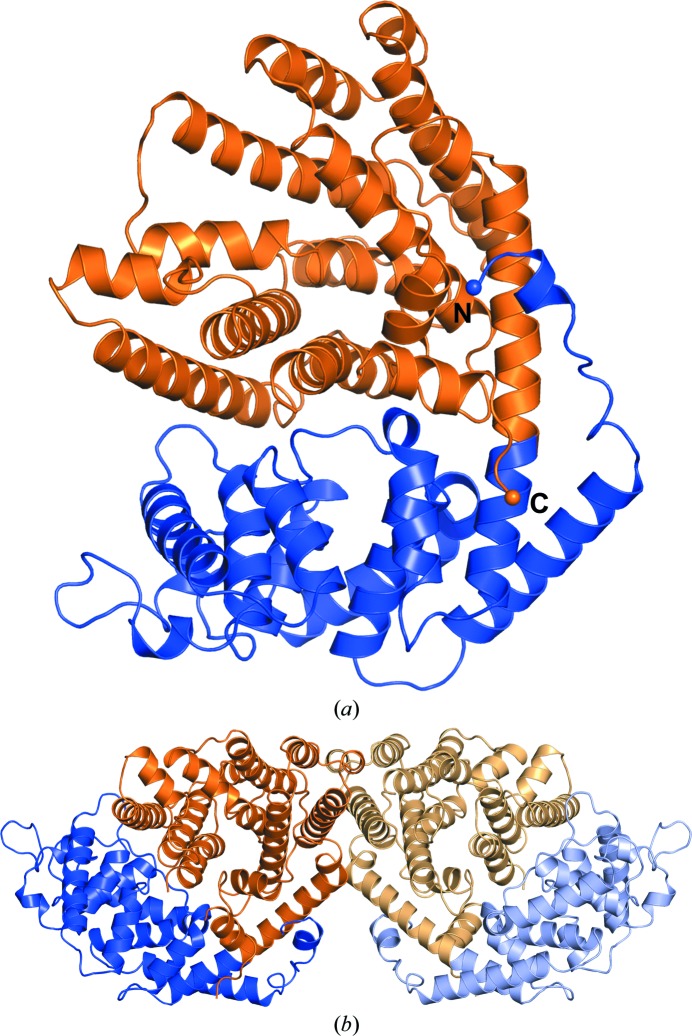
Figure 5.
Overall structure of TvTPS1, a two-domain protein. (a) Overall structure of a TvTPS1 monomer (chain B) with the N-terminal domain coloured blue and the C-terminal domain coloured orange. The N- and C-termini are depicted as spheres and labelled. (b) Dimer of TvTPS1 as observed in the asymmetric unit of the triclinic crystal with molecule A shown in lighter colours and molecule B in darker colours using the same domain-colouring scheme as in (a).

