
FIG. 2.
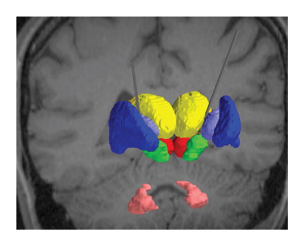
For the subset of individuals who underwent DBS of the STN, we created patient-specific computer models of DBS. Each model integrated into a common platform the patient imaging data, 3D subcortical nuclei, DBS electrode, and the VTA. The relevant nuclei were contoured and include the globus pallidus and putamen (blue), the thalami (yellow), the red nuclei (red), the STN (green), and the dentate nuclei (pink).