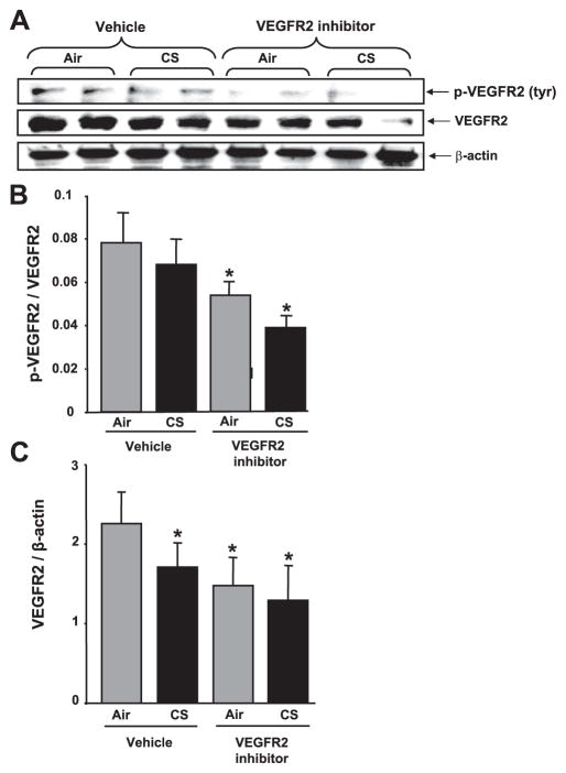
Figure 4.
VEGFR-2 levels and its phosphorylation were decreased in response to CS and/or VEGFR-2 inhibition in mouse lung. Soluble proteins from lung homogenates were electrophoresed in a 4 –15% gradient PAGE gel and electroblotted into nitro-cellulose membranes. A) p-VEGFR-2 and VEGFR-2 protein were measured using immunoblotting with rabbit anti-tyr(p)-VEGFR-2 (Clone 4G10-universal p-tyr antibody, Upstate Cell Signaling) and anti-VEGFR-2 primary antibodies (Cell Signaling), respectively. B) Histograms represent mean ± SE relative levels of p-VEGFR-2 (n=4). *P < 0.05 vs. both vehicle-treated air- and CS-exposed groups. C) Histograms represent mean ± SE relative VEGFR-2 expression in mouse lungs (n=4). *P < 0.05 vs. vehicle-treated air-exposed group.

