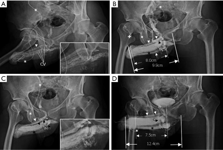
Figure 2.
Representative cavernosography of a 32-year-old male in the cavernosal vein group. (A) While the tip of a 19-gauge scalp needle (white asterisk) was positioned at the lateral aspect of the right corpus cavernosum, four independent channels of the cavernosal veins and the deep dorsal vein (white arrowhead) were visible; (B) four cavernosal veins (black arrowheads) and the deep dorsal vein (white arrowhead) were enhanced. The corpus spongiosum (black asterisk) was identifiable. Even two penile crura could be differentiated in addition to the septum; (C) those tissues were enhanced and the bulbo-urethral vein (white arrow) was visible; (D) the assembly of the deep dorsal vein (white arrowhead), a circumflex vein (black curved arrow) and the corpus spongiosum (black asterisk) were presented when an artificial erection was made.

