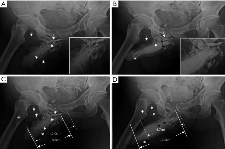
Figure 7.
Cavernosograms of a 45-year-old man in the deep dorsal vein group. (A) While the tip of a 19-gauge scalp needle (white asterisk) was positioned in the right corpus cavernosum, the deep dorsal vein (white curved arrow), the superficial dorsal vein (white arrows), and the cavernosal vein (white arrowhead) were shown after injection of the contrast medium. Several huge circumflex veins presented proximally; (B) those veins were enhanced while the right femoral vein (cross) was poorly opacified; (C) the venous complex of deep dorsal vein (white curved arrow), cavernosal veins (white arrowheads) and several superficial dorsal veins (white arrows) were clearly demonstrated. There was an extraordinarily complex venous plexus (black double arrows) composed of circumflex veins beneath the penile base; (D) the circumflex veins (black arrows) seemingly drained the cavernosal sinusoidal bloods to the right femoral vein (white cross) via several superficial dorsal veins (white arrows) when an artificial erection was induced.

