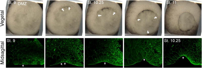
Figure 1.

Bottle cell formation as the first external sign of Xenopus gastrulation. Top, vegetal view of blastopore formation, with bottle cells forming initially in the dorsal marginal zone (DMZ), then laterally and ventrally to form the circular blastopore. Arrows mark the extent of apically constricting bottle cells. Bottom, midsagittal confocal images of bottle cells immunostained with α-tubulin antibody. Embryos are oriented apical down and animal to the right. Arrows point to center of blastopore invagination. St., stage. (Reprinted from Lee and Harland, 200720).
