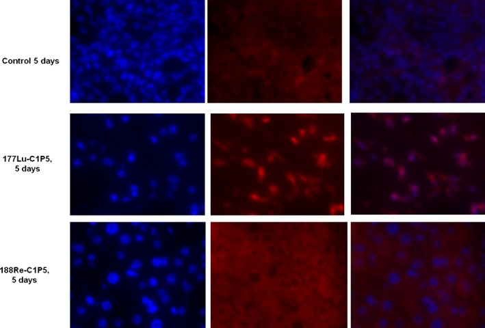
Figure 3.

Immunohistochemistry staining for gamma H2AX foci in 188Re‐C1P5 and 177Lu‐C1P5 tumors on Day 5 post radioimmunotherapy. First row—untreated controls, second row—177Lu‐C1P5, third row—188Re‐C1P5 treatment. First column—DAPI staining (blue), second column—gamma H2AX foci staining (red), third column—superimposition of DAPI and gamma H2AX staining.
