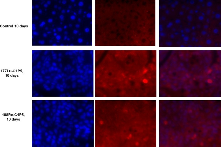
Figure 4.

Immunohistochemistry staining for gamma H2AX foci in 188Re‐C1P5 and 177Lu‐C1P5 tumors on Day 10 post radioimmunotherapy. First row—untreated controls, second row—177Lu‐C1P5, third row—188Re‐C1P5 treatment. First column—DAPI staining (blue), second column—gamma H2AX foci staining (red), third column—superimposition of DAPI and gamma H2AX staining.
