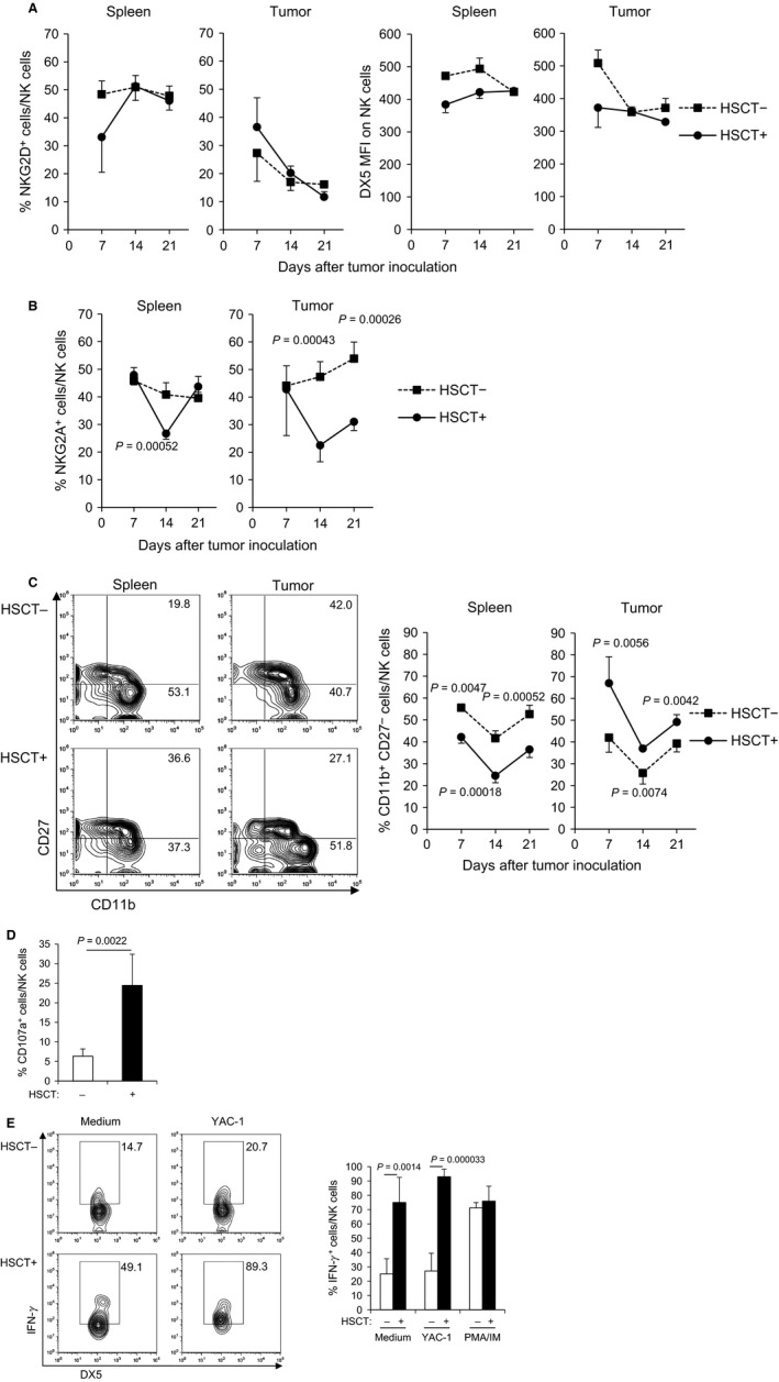
Figure 2.

NK cells in HSCT tumor showed mature phenotype with a low expression level of inhibitory receptor and were highly activated. (A) The expression of activating receptors on NK cells in the spleen and tumor after syngeneic HSCT. The tumors and spleens at days 7, 14, and 21 after tumor inoculation were harvested and the frequency of NKG2D+ cells within NK cells and MFI of DX5 on NK cells was analyzed by flow cytometry (n = 4). (B) The expression of inhibitory receptor on NK cells in the spleen and tumor after syngeneic HSCT. The frequency of NKG2A+ cells within NK cells was analyzed by flow cytometry (n = 4). (C) Maturity of NK cells in the spleen and tumor after syngeneic HSCT. The frequency of mature NK cells (CD11b+ CD27−) within NK cells was analyzed by flow cytometry (n = 4). Representative flow cytometry plots of spleen and tumor at day 21 after tumor inoculation are shown (left panel). (D) Cytotoxicity of NK cells in tumor at day 14 after syngeneic HSCT. The percentage of CD107a+ cells within NK cells was analyzed by flow cytometry (n = 4). (E) Cytokine production of NK cells in tumor at day 14 after syngeneic HSCT. TILs isolated from tumors were restimulated with YAC‐1 tumor cells ex vivo. Then, IFN‐γ intracellular cytokine staining was performed and the frequency of IFN‐γ + NK cells was analyzed by flow cytometry (n = 4). Representative flow cytometry plots are shown (left panel). NK, natural killer; HSCT, hematopoietic stem cell transplantation; MFI, mean fluorescence intensity; TILs, tumor‐infiltrating lymphocytes.
