
Figure 4.
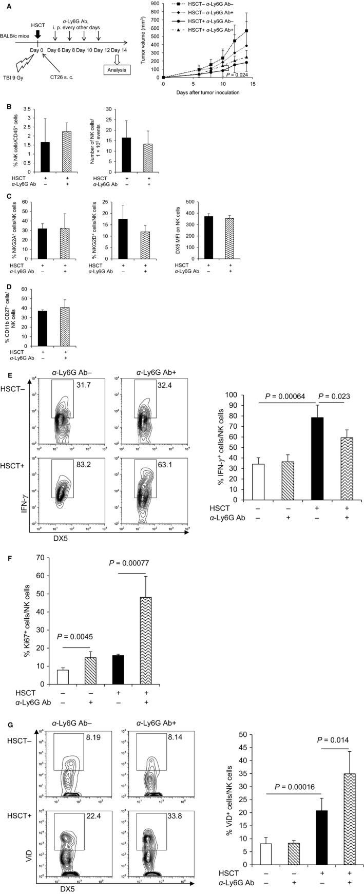
Neutrophil depletion attenuated activation of NK cells in HSCT tumor. Tumors were harvested at day 14 after syngeneic HSCT, and TILs were isolated from tumors and analyzed by flow cytometry. (A) Tumor growth suppression in the neutrophil‐depleted mice. Transplanted or non‐treated BALB/c mice were intraperitoneally injected with anti‐Ly6G antibody every other day after tumor inoculation to deplete neutrophils (number of animals per each group: n = 6–8). The tumor volume was measured at the indicated days after tumor inoculation. Data are shown as mean ± SD. (B) Frequency and number of NK cells in HSCT tumor after syngeneic HSCT with neutrophil depletion. The frequency of NK cells and number of NK cells within fixed number of tumor cells (1 × 10⁵) were analyzed (n = 4). (C) The expression of activating and inhibitory receptors on NK cells in HSCT tumor with neutrophil depletion. The frequency of NKG2A+ cells (left panel) and NKG2D+ cells (middle panel) within NK cells, and MFI of DX5 on NK cells (right panel) were analyzed (n = 4). (D) Maturity of NK cells in HSCT tumor with neutrophil depletion. The frequency of mature NK cells (CD11b+ CD27−) within NK cells was analyzed (n = 4). (E) IFN‐γ production of NK cells in HSCT tumor with neutrophil depletion. TILs isolated from tumors were restimulated with YAC‐1 tumor cells ex vivo. Then, IFN‐γ intracellular cytokine staining was performed and the frequency of IFN‐γ + NK cells was analyzed by flow cytometry (n = 3–4). Representative flow cytometry plots are shown (left panel). (F) Ki67+ cells within NK cells in HSCT tumor with neutrophil depletion. Ki67 intracellular staining was performed and the frequency of Ki67+ NK cells was analyzed (n = 4). (G) Dead cells within NK cells in HSCT tumor with neutrophil depletion. Dead cell staining was performed and the percentage of ViD+ dead NK cells was analyzed (n = 4). Representative flow cytometry plots are shown (left panel). NK, natural killer; HSCT, hematopoietic stem cell transplantation; TILs, tumor‐infiltrating lymphocytes; MFI, mean fluorescence intensity; ViD, viability dye.