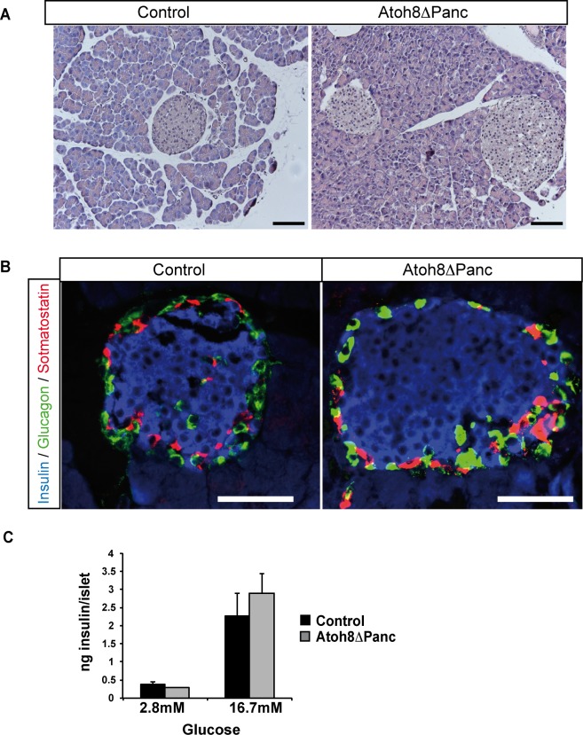
Fig 4. Pancreas morphology and islet insulin secretion in 36-week old Atoh8 Δpanc mice.
(A) Hematoxilin-eosin staining showed no apparent differences in pancreas gross morphology between 36-wk-old Atoh8 Δpanc mice and controls. Scale bars 100 μM (B) Immunofluorescence staining for insulin (blue), glucagon (green) and somatostatin (red) on pancreas sections from 36-wk-old Atoh8 Δpanc mice and controls. Representative images demonstrate similar islet cell organization between mutants and controls. Scale bars 50 μM (C) Glucose-induced insulin secretion in isolated islets from 36-wk-old Atoh8 Δpanc mice and controls. n = 3 mice per genotype (3–4 independent islet batches per animal); mean ± SE.

