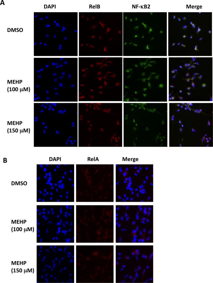
Fig 2. MEPH exposure increases nuclear RelB/p52.
Primary term cytotrophoblast were exposed to MEHP for 24 hours at concentrations as indicated with DMSO as the vehicle control. Immunofluorescence (IF) staining was performed for direct visualization of intracellular distribution of RelB/p52 (A) and RelA (B), as detailed in the Materials and Methods. These images represent data obtained from three independent placentas. Original magnification, 200Χ.

