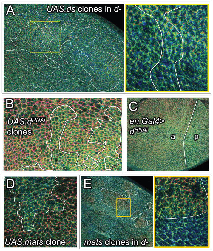
Figure 5. Ft/Ds signaling acts via Dachs to inhibit or reverse the “closed” to “open” transition of Wts mediated by Mats.
A) Q ratio image of a d− wing disc containing numerous, large UAS.ds over-expressing clones: the yellow box includes a clone located within the prospective wing, shown at higher magnification underneath (Q ratio scale for A–E as in Fig. 2). In contrast to wt discs, d− discs appear predominantly green and clones of UAS.ds over-expressing cells in such discs fail to induce an increase in Q in surrounding cells (compare with Fig. 4A), indicating general occupancy of the CWT sensor in the “open” state, irrespective of Ft/Ds signaling.
B) Q ratio image of wing tissue containing clones of cells in which Dachs activity has been knocked down by UAS.dRNAi expression. Clones appear green relative to the surround. Similar results were obtained for clones of homozygous d− cells, although it was necessary either to over-express Yki in such clones, or to generate them in wing discs with reduced ex gene function (Fig. S7), to allow the d− cells to grow similarly to surrounding cells that retain normal d function.
C) Q ratio image of a wing disc in which Dachs activity has been knocked down selectively in the posterior (p) compartment by expressing UAS.dRNAi under en.Gal4/UAS control. The posterior compartment (p), which is abnormally small, is predominantly green/blue. In contrast, the anterior (a) compartment shows the normal central-to-peripheral, green-to-red shift.
D) Q ratio image of proximal wing tissue containing a clone of UAS.mats over-expressing cells. The clone appears green relative to the surround, indicating increased occupancy of the CWT sensor in the “open” state.
E) Q ratio image of a d− wing disc with several matse03077 clones (some outlined in white); a portion of one such clone, boxed in yellow, is shown at higher magnification to the right. The clones appear red, in contrast to the surround, which appears blue-green, indicating that occupancy of the CWT sensor in the “open” state in d− cells depends on Mats.

