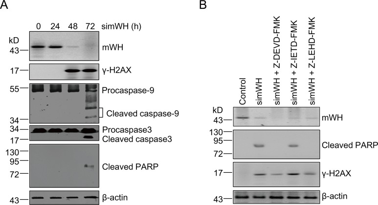
Fig 3. DNA damage precedes apoptosis after mWH depletion in mouse JB6 cells.
(A) Time course of DNA damage and apoptosis following mWH depletion. Samples were collected for analysis following mWH knockdown at 0, 24, 48, and 72 h. DNA damage (γ-H2AX signals) was detected at 48 h after siRNA treatment. Apoptotic signals including cleavage of Procaspase-9, Procaspase-3, and PARP, were detected only after 72 h of treatment. (B) Inhibitors of Caspases-9 and -3, but not for Caspase-8, block apoptosis ensuing depletion of mWH. However, DNA damage signals were unaffected and present under these conditions. Cells were co-cultured with simWH and different Caspase-specific inhibitors (final concentrations of 2 μM in the culture medium) including Z-DEVD-FMK (Caspase-3), Z-IETD-FMK (Caspase-8), and Z-LEHD-FMK (Caspase-9). After 72 h of treatment, samples were collected and processed for western blot.

