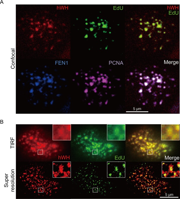
Fig 7. Localization of hWH at the site of DNA replication determined by confocal (A) and super-resolution microscopy (B).
(A) HCT116 p53+/+ cells were stained with rabbit anti-hWH antibody (red), mouse anti-FEN1 antibody (blue), goat anti-PCNA (purple), and the nascent DNA replication marker EdU (green). The merged images of hWH plus EdU, or all four signals, are in the upper-right and lower-right panels, respectively. (B) HCT116 p53+/+ cells were stained with rabbit anti-hWH antibody (red) and EdU (green). The images were obtained by TIRF microscopy and super-resolution microscopy. The boxed areas are shown at higher magnification.

