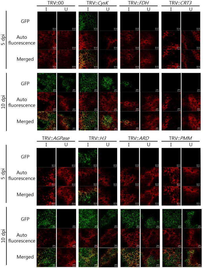
Fig 4. Effects of gene silencing on CMV-P1-GFP infection in N. benthamiana.
GFP fluorescence was observed at 5 dpi and 10 dpi in VIGS plants, and TRV::00 plants were used as a positive control. Images on the left are optical sections of the inoculated leaves, and those on the right are optical sections in the upper leaves. Images top to bottom are GFP, autofluorescence, and merged images, respectively. The green fluorescence signal indicates CMV-P1 expressing GFP, and red fluorescence signal indicates chloroplasts. Scale bars = 50 μm.

