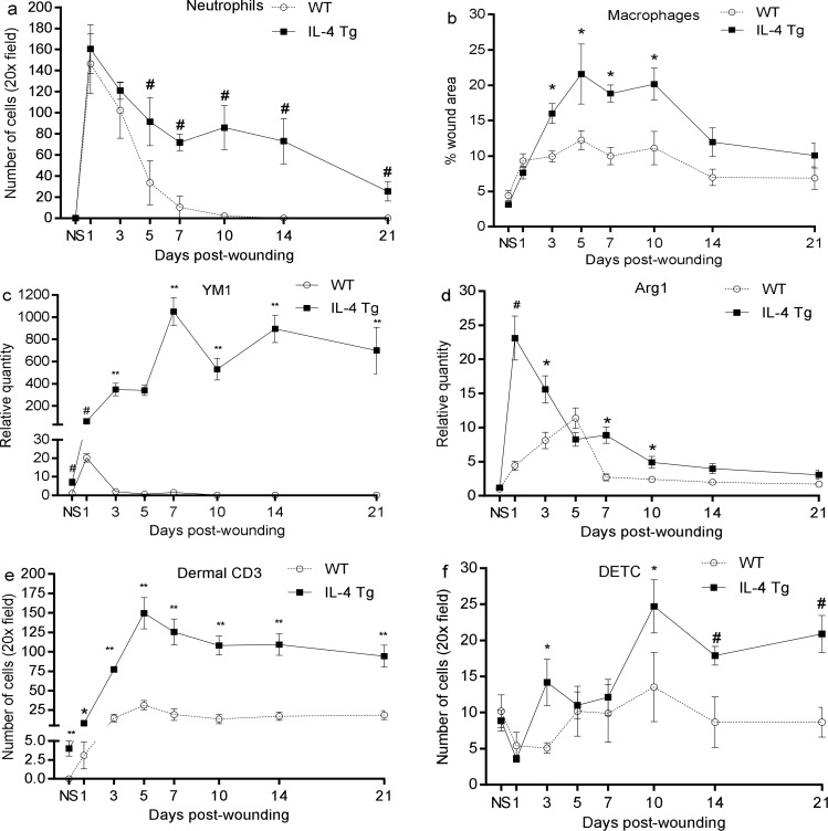
Fig 3. Injury induces robust inflammatory cell infiltration in wounds of IL-4 Tg mice.
Three mm full thickness excisional wounds were made on dorsal skin of IL-4 Tg and WT mice. At various time points, frozen sections were prepared for neutrophil (Gr-1), macrophage (CD68), CD3, and DETC staining. Positively stained cells in the wounds and wound margins were counted and the average number per 20x field was calculated. a and b) Time course of the number of neutrophils and macrophages, respectively. c and d) YM1 and Arg1 mRNA expression in wounds respectively. e and f) Time course of the number of dermal CD3+ lymphocytes and DETC, respectively. * p<0.05, # p<0.01, and ** p<0.001compared to WT at the same time point, respectively. NS: normal or unwounded skin. The number of mice used at each time point was 5.

