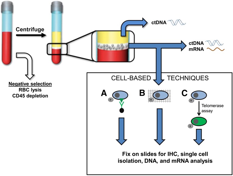
Figure 1.
Overview of circulating tumor cell (CTC), ctDNA, and mRNA isolation techniques. Negative selection applied to whole blood removes RBCs and CD-45-expressing leukocytes. After separation of blood components, ctDNA can be extracted from plasma (yellow layer). The mononuclear layer (white layer) can be used for ctDNA, mRNA, or CTC extraction (illustrated in the box labeled Cell-Based Techniques). CTC detection and isolation can result in downstream IHC analysis, single cell isolation, and DNA and mRNA analysis. Lane A: Surface-marker dependent detection methods use antibodies against melanoma-specific surface marker antigens. Antibodies may be linked to ferrous beads for magnetic pull-down or fluorescent proteins for visualization. White blood cells (WBCs) are represented in the illustration by a small gray cell. Lane B: Surface-marker independent detection methods whereby CTCs may be captured along with WBCs on a fine porous barrier. This is termed the ISET, or isolation by size of epithelial tumor cells, method. Lane C: A telomerase live-cell assay applied to CTCs and WBCs causes cells with elevated telomerase promoter activity to produce high levels of green fluorescent protein.
Abbreviations: ctDNA, circulating tumor DNA; IHC, immunohistochemistry; RBC, red blood cell.

