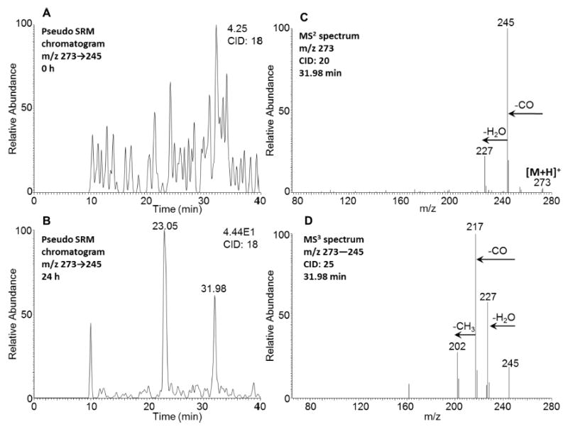
Figure 6.

Detection of 5-MC-1,2-dione in human HepG2 cells. (A) Extracted ion chromatogram of pseudo SRM transition at 0 h. (B) Extracted ion chromatogram of pseudo SRM transition at 24 h. (C) MS2 spectrum of the peak at 31.98 min. (D) MS3 spectrum of the peak at 31.98 min. The samples were prepared as described in the caption to Figure 1 and were subsequently analyzed on an ion-trap LC-MS/MS. Another peak with a retention time of 23.05 min and with relatively high polarity could be an isomer of O-monosulfonated-5-MC-catechol, which could undergo cleavage of the sulfate conjugate in the mass spectrometer followed by auto-oxidation and thus result in the detection of quinone instead.
