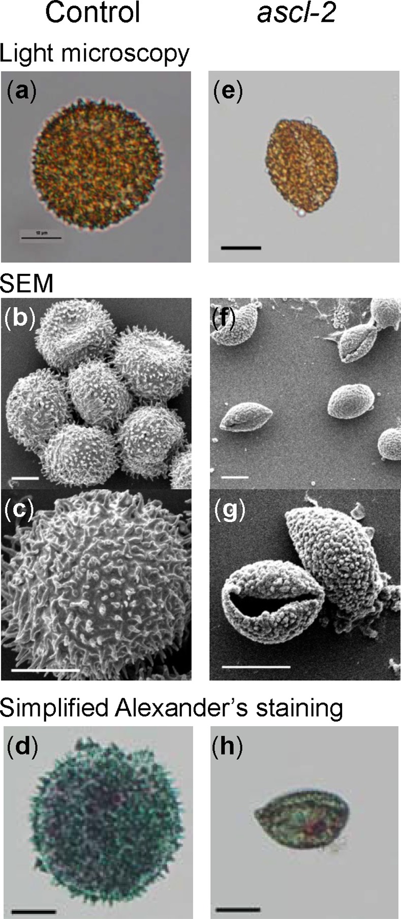
Fig 6. Morphological comparison of orange stage pabB4 control and ascl-2 spores.

Images of typical spores isolated from mature orange sporophytes were acquired using light microscopy (a,e) and SEM (b,c,f,g). Spores from the control (a–c) and ascl-2 (e–g) are shown. Light microscopy images of control (d) and ascl-2 (h) spores after treatment with simplified Alexander’s stain are also shown. Scale bars = 10 μm.
