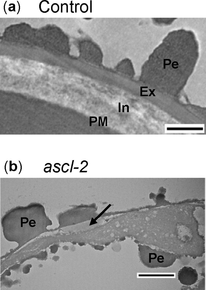
Fig 7. Transmission electron micrographs of pabB4 control and ascl-2 spores.

Cross sections of spores from mature orange control (a) and ascl-2 (b) sporophytes were examined with TEM. An amorphous layer found below much of the perine is indicated by an arrow in (b). Ex, exine; In, intine; Pe, perine; PM, plasma membrane. Scale bars = 500 nm.
