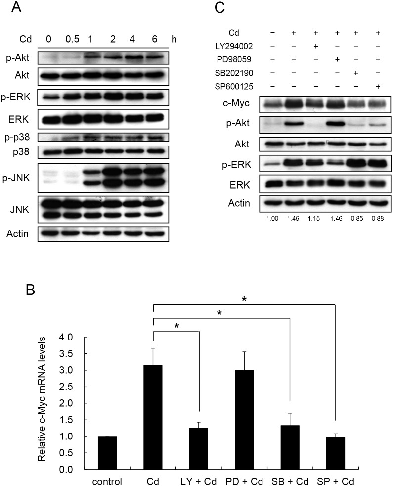
Fig 3. Cd activates PI3K, p38 and JNK signaling factors to increase c-Myc mRNA and protein.
HepG2 cells were treated with 5 μM Cd for 6 hours. Samples were removed at various time intervals and the phosphorylation of the signaling factors was determined by Western blotting. (B) Cells were treated with 50 μM of inhibitor for PI3K (LY294002), ERK (PD98059), p38 (SB202190) and JNK (SP600125) 1 h prior to the addition of 5 μM Cd. Cells were cultured for additional 4 h. Total RNA was extracted and the c-Myc mRNA was quantified by real-time PCR. Each value represents a mean ± standard deviation of three samples. Asterisks (*) indicate significant differences (p < 0.05) between the paired samples. (C) Cellular c-Myc after various inhibitor treatments as in (B) was analyzed by Western blotting. Phospho-Akt was detected by antibodies specific for the modification at Ser473. Phosphate proteins are designated with ‘p-‘ in front of the protein name. LY: LY294002; PD: PD98059; SB: SB202190; SP: SP600125. Numbers underneath the Western blotting represent the amount of c-Myc relative to that of the loading control (actin).

