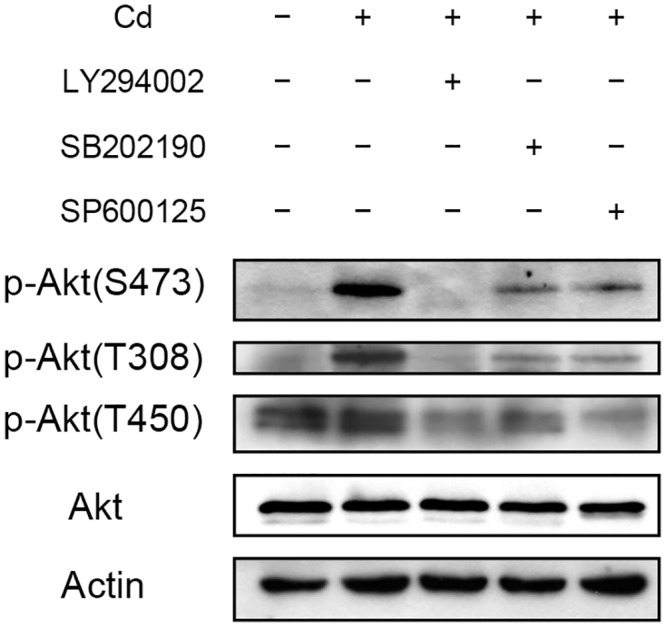
Fig 5. Effect of Cd on Akt phosphorylaion at different sites.

HepG2 cells were treated with 50 μM of PI3K ((LY294002), p38 (SB202190) or JNK (SP600125) inhibitor 1 h prior to the addition of 5 μM Cd. Cells were cultured for additional 4 h then harvested and prepared for Western blotting. Antibodies raised specifically against phosphorylation at Thr308 [p-Akt(T308)], Thr450 [p-Akt(T450)] or Ser473 [p-Akt(S473)] of Akt were used for the analysis. Actin was used as a loading control for immunoblotting.
