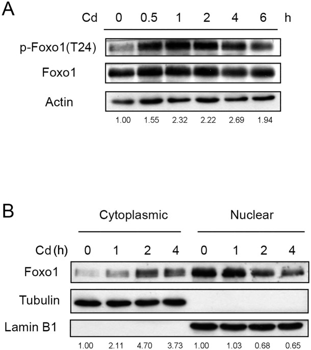
Fig 6. Cd stimulates the phosphorylation of Foxo1 and translocations the protein from the nucleus to the cytoplasm.
(A) HepG2 cells were treated with 5 μM Cd for various time intervals and the levels of Foxo1 and phospho-Foxo1 [p-Foxo1(T24)] were analyzed by Western blotting. Numbers underneath the Western blotting represent the amount of phospho-Foxo1 relative to that of the loading control (actin). (B) Cells were treated with 5 μM Cd for various time intervals. Cells were harvested and cytosolic and nuclear fractions were prepared for Western blotting. Numbers underneath the Western blotting represent the amount of Foxo1 relative to that of the loading control (tubulin for cytoplasmic proteins and lamin B1 for nuclear proteins).

