Fig. 19.

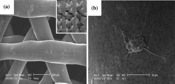
SEM images of PCL/TCP composite scaffolds obtained from FDM: a structure of top view with inset of cross-sectional view; and b osteoblast cells attached on the scaffold surface (Zhou et al. 2007)

SEM images of PCL/TCP composite scaffolds obtained from FDM: a structure of top view with inset of cross-sectional view; and b osteoblast cells attached on the scaffold surface (Zhou et al. 2007)