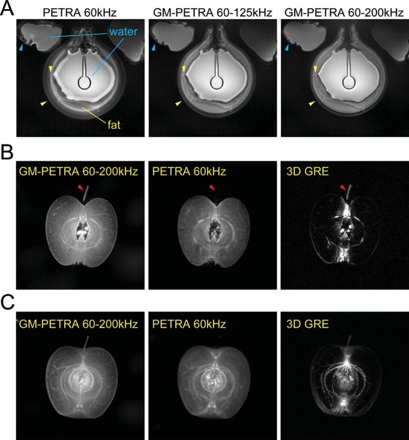
Figure 3.
(A) Improvement of the off-resonance artifacts as a result of chemical shift (fat-water) and susceptibility differences in a phantom experiment with GM-PETRA. Duplicated edges around fat regions became thinner as the GM increased; small gaps between fat compartments were visualized better in images from higher bandwidth (yellow arrowheads). Image blurring and distortion around the edges of water compartments also improved along with an increase of the bandwidth (light blue arrowheads). PETRA 120 kHz showed similar image blurring artifacts to GM-PETRA 60–125 kHz (data not shown), which is consistent with the simulation results shown in Figure 2. (B) Image blurring as a result of short T2* decay and/or off-resonance demonstrated in an apple imaging. GM-PETRA allowed finer structures in the mesocarp (flesh) to be appreciated compared to PETRA; these structures were completely invisible in 3D GRE because of their very short T2* values. Although the calyx had a relatively long T2* (which makes it visible with 3D GRE), it was completely blurred out with PETRA because of strong susceptibility effects (red arrowheads). (C) An MIP image from GM-PETRA clearly allows fine structures in the mesocarp to be appreciated, including fibers compared to the other 2 acquisition methods.

