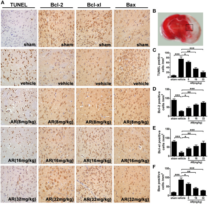
Figure 2.
The effect of AR on neurons apoptosis and bcl-2, bcl-xl, and bax expressions in the penumbral area. The vehicle group and AR groups underwent MCAO while the sham group underwent the same surgical procedure without the filament inserted. After MCAO performed, rats in AR groups were intraperitoneally administrated with AR (8, 16, and 32 mg/kg) for 3 days. (A) Representative photographs of TUNEL staining, immunohistochemical staining of bcl-2, bcl-xl, and bax in the cortex of MCAO rat. (B) The black square indicates the penumbral area. (C) TUNEL staining was analyzed by Image Pro Plus, and each datum was presented as TUNEL positive cells numbers in per mm2 within five random independent view fields. Quantification of the levels of bcl-2 (D), bcl-xl (E), and bax (F) expressions was presented as immunohistochemical positive cells numbers in per mm2 within five random independent view fields. The values were presented as mean ± SD (n = 8). *P < 0.05; **P < 0.01; ***P < 0.001.

